Nano-MIPs
|
In collaboration with Prof. Karsten Haupt, UTC, France |
|
The technique of molecular imprinting allows for the preparation of synthetic polymers with specific binding sites for a target molecule. This can be achieved if the target is present during the polymerization process, thus acting as a molecular template. Monomers carrying certain functional groups are arranged around the template through either noncovalent or covalent interactions. Following polymerization with a high degree of cross-linking, the functional groups become fixed in defined positions by the polymer network. Subsequent removal of the template by solvent extraction or chemical cleavage leaves cavities that are complementary to the template in terms of size, shape and arrangement of the functional groups. These highly specific receptor sites are capable of rebinding the target molecule with high specificity, sometimes comparable to that of antibodies. Molecularly imprinted polymers have therefore been named "antibody mimics". It has been shown that they can be substituted for biological receptors in certain formats of immunoassays and biosensors. In an attempt to use this technique to realize nano-arrays of molecularly imprinted polymers (MIPs), we deposit the pre-polymerization mixture using NFP, and subsequently (UV) polymerize the resulting nanostructures, much like in our work on microlenses. |
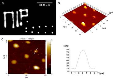 When the template is a fluorescent molecule (fluorescein, in this case), the array and other written features are directly observable with a fluorescence microscope. |
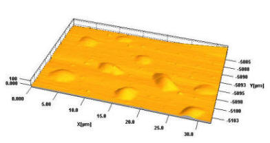 An example of less than 100 nm thick dots of MIPs |
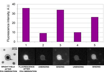
A fluorescent template molecule enables us to directly observe binding and releasing of the analyte, by observation of the fluorescence intensity, in a flow cell mounted on the stage of the fluorescence microscope.
|
|
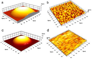 Single dots of different compositions. The one in a and the zoom in showed in b is more porous (thus the cavities are more accessible to the analyte) than the one in c and (zoomed image in) d. |
Unbinding and rebinding experiment, including control (non-imprinted) dots. The four small dots, surrounding the large central one, are non-imprinted. The different size is intentional, to enable easy distinguishing of the imprinted and non-imprinted dots. It is obvious that the imprinted dot binds (and releases) fluorescein, while the four non-imprinted ones do not bind it. |
|
|
|