Measurement of forces applied by cilia with AFM
|
In collaboration with Prof. Zvi Priel
|
|
Cilia are motile protrusions from a myriad of specialized cells. Their role in most systems is biological transport, from propulsion of protozoa in water to removal of foreign matter in mammalian trachea and ingestion assistance in frog’s esophagus. Mucus propelling cilia are 5-7 mm long, 0.2 mm thick, densely packed (~ 1 mm interciliary spacing) and continuously beating. |
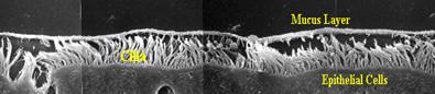 SEM of mucus propelling cilia |
||
 Commonly accepted beat cycle of a cilium. Positions 1-9 : fast effective stroke. Positions 9 – 12: slow recovery stroke (on an inclined plane) |
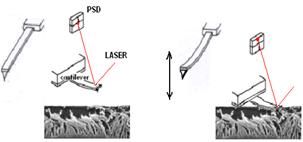 An AFM tip is dipped in a field of beating cilia. Cilia hitting the tip are causing it to rock, and its motion is detected by the laser beam focused on its back. |
||
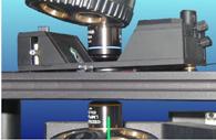 |
The experimental setup consists of a flat-scanner AFM, leaving a clear optical path for two optical microscopes. Simultaneously with the AFM measurement, light is shone on the ciliary cells through one objective, and collected through the other objective, after passing through the beating cilia. The modulated light is converted with a PMT and sampled with a digital card. |
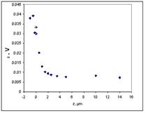 |
The force sensed by the AFM increases with the decrease of probe-sample separation. The deeper the probe penetrates the more cilia hit it. The curve is starting to bend up at around 5 mm, which correlates nicely with the known length of the cilia. Our measurements show: Applied force per cilium 710 ± 230 pN |
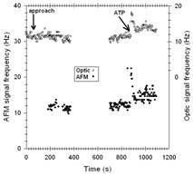 |
Simultaneous optical measurement assures that no mechanical stimulation is induced by the AFM probe. The frequency before and after AFM probe approach are identical. Stimulation of beat frequency with extracellular ATP causes identical frequency increase in both signals. |
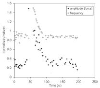 |
AFM measurement showing the amplitude and frequency of the AFM tip, as the cilia are stimulated with extracellular ATP. The relationship frequency-amplitude is linear: the log-log plot has a slope of 1. This indicates that the length of the effective stroke arc remains unchanged.  |
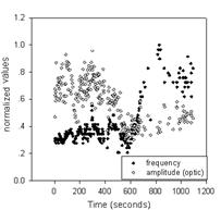 Optical Measurement |
 AFM measurement |
During a stimulation event, the frequency is increasing as reported by both optical and AFM signal. The amplitude of the AFM signal is increasing, while that of the optical signal is decreasing. This indicates that while the arc length of the effective stroke does not change, the plane on which the recovery stroke is performed, becomes more perpendicular to the plane of the cell membrane |
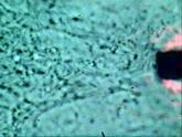 A field of beating cilia and the AFM tip, as viewed through the optical microscope. (click the image to play) |