Total Internal Reflection Fluorescence Microscopy
Total internal reflection fluorescence microscopy (TIRFM) employs the unique properties of an induced evanescent wave to selectively illuminate and excite fluorophores in a restricted specimen region immediately adjacent to a glass-water (or glass-buffer) interface.
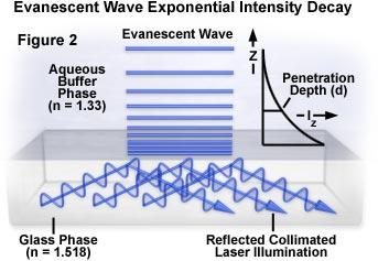
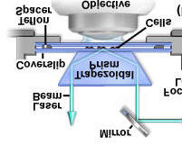
The basic concept of TIRFM is simple, requiring only an excitation light beam traveling at a high incident angle through the solid glass coverslip or plastic tissue culture container, where the cells adhere. Refractive index differences between the glass and water phases regulate how light is refracted or reflected at the interface as a function of incident angle. At a specific critical angle, the beam of light is totally reflected from the glass/water interface, rather than passing through and refracting in accordance with Snell's Law. The reflection generates a very thin electromagnetic field (usually less than 200 nanometers) in the aqueous medium, which has an identical frequency to that of the incident light. This field, called the evanescent wave or field, undergoes exponential intensity decay with increasing distance from the surface.
 q(c) = sin-1n(2)/n(1) = sin-1n(2)
(if n=1, i.e. air)
q(c) = sin-1n(2)/n(1) = sin-1n(2)
(if n=1, i.e. air)
I(z) = I(o)e-z/d
where: d = l(o)/4p (n(1)2sin2q(1) - n(2)2)-1/2
Image courtesy of www.microscopy.fsu.edu
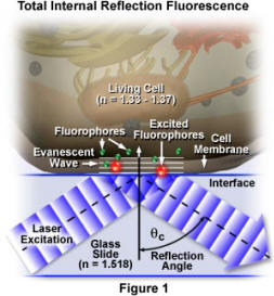
Image courtesy of www.microscopy.fsu.edu
mage courtesy of www.microscopy.fsu.edu
Original image, in legible orientation
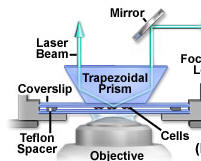
Image courtesy of www.microscopy.fsu.edu