Building 63 room 20
Tel:
08-6477798
Head: Ms. Roxana Golan - CV
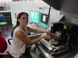
The Minreva laboratory shares the space and equipment with the Ilse
Katz center for Nanotechnology (http://www.bgu.ac.il/nanocenter/)
enabling the utilization of a variety of SPM techniques.
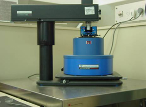
The Veeco (formerly TM-Park) CP Research is a closed-loop commercial
scanning probe microscope. It is equipped with an optical imaging
system, with a large SPM scanner for scans up to 100 mm and a high
resolution SPM scanner for scans smaller than 5 mm. Available modes
include contact, intermittent contact, lateral force, electrical,
magnetic and scanning capacitance modes. Imaging is possible in air
as well as under liquid.
Highlights - research projects
carried out in the AFM lab
Structural characterization of
polydiacetylene Langmuir films in the presence of various divalent
subphase cations (A. Berman, Y. Golan, Materials
Eng http://www.bgu.ac.il/materials/hp/index.htm)
The linear strand morphology of polydiacetylene Langmuir
film was characterized and the morphological effects of various metal
ions were studied. These films are used as templates for the deposition
of metal-sulfide semiconductor nanocrystals that are organized along
the linear strands of the polymer.
Figure 1: AFM
images (4 ?m) of PCDA monomers compressed and polymerized on progressively
increased concentration of Cd2+ ions in subphase. This causes the
morphology of the films to change from large domains with linear strand
morphology to much smaller ones which do not exhibit any strand morphology.
Dynamic Behavior of DPPC:POPG and Commercial
Lung Surfactants
(M. Gordon, R. Granek, A. Berman, Biotechnology Eng.)
Pulmonary surfactant is a fundamental substance in the mechanics
of the breathing process. Lung surfactant (LS) spread at the air-liquid
interface of the alveoli is described as a monolayer of phospholipids
and protein molecules. Its main function is to prevent collapse of
the alveoli during expiration by reducing the surface tension at the
air liquid interface of the lungs. The dynamic behavior of LS and
DPPC-POPG 4:1 mixture is studied on Langmuir film balance (Langmuir
trough) and the film organization is analyzed in-situ with Brewster
Angle Microscope and ex-situ with fluorescent microscopy (FM). AFM
is a major tool for following the morphological changes (with angstrom
resolution in the z-direction) that take place in these surfactant
films during compression and expansion.
Figure 2:
AFM images of exogenous LS indicating on fingers formation at the
center of LC domains.
Morphology evolution in nanocrystalline PbSe
and PbS chemically deposited on GaAs (M. Shandalov, A. Osherov
and Y. Golan (http://www.bgu.ac.il/materials/hp/index.htm),
Materials Eng.)
PbSe thin films are deposited using a simple and inexpensive deposition
technique. The shape, size and lateral distribution of PbSe nanocrystals
are studied using AFM. Deposition on GaAs substrates is compared to
deposition on Si under the same conditions. In another sub-project,
nano-PbSe is deposited on GaAs(100) onto which sub-?m trenches are
micropatterned using laser interference lithography. Recently PbS
films prepared using the same technique.
Figure 3. AFM
Images of micropatterned GaAs surfaces onto which nanocrystalline
PbSe was grown using chemical solution deposition.
Adhesion studies of biolubricants
extracted from a species of microalga on mica surfaces (S.
Arad (arad@bgu.ac.il), Biotechnology
Eng., Y. Golan, Materials Eng. (http://www.bgu.ac.il/materials/hp/index.htm),
L. Rappaport, HAIT)
The increase in friction forces with time in biolubricated
contacts often occurs when the biolubricant is squeezed out of the
contact. In order to compare the adhesion of biolubricants extracted
from microalga to hyaluronic acid (a conventionally used bio lubricant),
the two materials are applied onto mica surfaces, thoroughly washed
and then imaged with AFM. The results show that the biolubricants
extracted from microalga remain adsorbed to the surface while hyaluronic
acid is washed out.
Figure 4. AFM Images of mica surfaces
lubricated with polysaccharide solution, rinsed and dried. (a) 2%
polysaccharide, 5-mm scan, z-scale 10 nm, (b) 0.1% polysaccharide,
5-mm scan, z-scale 10 nm; (c) 0.02% polysaccharide, 3-mm scan, z-scale
3nm.
Characterization of PbTe semiconductor films
(R. Kreisman, Z. Dashevsky, Materials Eng.)
Grain boundaries in PbTe thin films serve as potential barriers for
charge carriers, and therefore highly affect the current and photo
current behavior of these films.
The effect of grain boundary density and properties on the electrical
and photoelectrical behavior of PbTe thin films is examined in this
project. The density and local topography of the films is examined
using AFM.
Figure 5: AFM Image showing the
grain structure observed in the surface PbTe.
Optimization of cleaning and restart sequence
of ultrafiltration membranes treatment process to enhance removal
of nature organic material (E. Arkhangerlsky, V. Gitis, Biotechnology
Eng).
Penetration of contaminants such as E. coli bacteria, T4 virus and
DNA proteins into membrane as a function of pore size is investigated.
Figure 6:
AFM Image showing the T4 virus in material filtered through an ultrafiltration
membrane.
Membranes for separation technology
(S. Freger, The Institute for Applied Research, BGU (http://www.bgu.ac.il/IAR/index.php)
Structural characterization of membranes used for micro-separation
technologies was carried out. The images below show an AFM scan of
an ultrathin active layer of polyamide composite membranes (skin).
Thickness measurements and morphological characterization of the interface
between the polyamide and support layers were carried out. AFM was
employed for assessment of the thickness changes in various liquid
environments, providing important information for the analysis of
permeability of the composite membranes of this type.
Figure 7: AFM
images showing the ultrathin active layer in a polyamide composite
membrane in air (left) and under water (right).
Force measurements of chemically modified
surfaces using the AFM (S. Botbol, J. Cohen, S. Efrima)
Forces between bare and chemically modified Si3N4 tips and glass
substrates that were coated with self-assembled monolayers were measured.
The AFM force measurements and microscopy images were coupled with
macroscopic contact angle measurements. A total of six different interaction
pairs were studied, including sulfonate and thioacetate self assembled
monolayers on glass surfaces.
Figure 8. Force curves of (a) Glass
modified with thioacetate self-assembled monolayer. (b) Force-distance
curve with sulphonate self-assembled monolayer. Schematic illustrations
of the two types of monolayers are shown below the force curves.
Inclusion of DNA in Liposomes (M. Zaccai
and Z. Weisman, The Institute for Applied Research, BGU)
Encapsulation of DNA in liposome structures was investigated. DNA
plasmid (6.5 kb) cut once by restriction enzyme is shown before inclusion
( left image). Note the DNA conformation. The shape and size of the
newly formed DNA -encapsulated vermonia liposomes were obtained from
the image on the right.